Cruciate Repair Surgery
Cranial Cruciate Ligament
(CCL) Tears in Dogs
The most common cause of hind-limb lameness in dogs, that is effectively treated with surgery for your dog to regain mobility and function in the affected leg.
Family Vet offer a range of common surgical solutions with fast recovery rates and long-term comfort and mobility for your dog.
What is a Cranial Cruciate Ligament (CCL) Tear?
The cranial cruciate ligament (CrCL or CCL) is the main stabilising ligament inside your dog’s knee (stifle) joint. In humans it is revered to as an ACL (Anterior Cruciate ligament). This ligament prevents the thigh bone (femur) from sliding down the lower leg (tibia) during movement. It also works together with other ligaments around the knee joint to prevent excessive rotation and dynamic stability when your dog walks or do exercise.
Once torn, the knee becomes unstable and abnormal. This instability causes the knee to rotate at abnormal angles and move excessively, which causes a lot of friction on the cushions (meniscus) inside the joint. These C shaped cartilages are often damaged or torn, especially in more unstable joints.
Unlike in people, most cruciate ligament ruptures in dogs are not caused by sudden trauma. In dogs, the ligament slowly degenerates over time due to multiple factors such as genetic factors, breed, conformation, obesity, and age. Most partial tears will progress to complete ruptures. The cruciate ruptures in a dog’s joint that is already affected by arthritis and show abnormal changes and inflammation.
Once the cruciate ligament is torn, the knee of the dog becomes unstable, which leads to pain, inflammation, and eventually arthritis, especially if left untreated.
The CCL prevents the tibia from sliding forward relative to the femur, once torn the two bones move more and grinds on the meniscus (in blue).
Image copyright Rebecca Jones (Diagram for educational purposes courtesy of Canine Cruciate Registry)
Common Causes & Risk Factors: Why did my dog rupture its Cranial Cruciate Ligament?
👵
Degeneration
Degeneration occurs commonly over months to years which causes the ligament ot weaken and eventually rupture. Some ruptures occur without any known trauma; it may even happen from just standing up! Owner often report that they suddenly noticed their dog lifting the leg up, or not standing on it, whilst limping at a gait.
This degeneration happens simultaneously in both joints and is the main reason why many dogs that rupture their one cruciate ligament, will subsequently rupture the other side within a few months to years.
🐕
Breed predispositions & Genetics
Labrador Retrievers, Rottweilers, Newfoundlands, Staffordshire Terriers, and many large breeds are reported to have an increased risk of rupturing their cruciate ligaments. The strength of the cruciate ligament differs between breeds with some breeds having a much stronger ligament as compared to other breeds. Smaller breed dogs such as Miniature and tea cup poodles, Yorkshire Terriers and others are often found with ruptures as well, although conformation and presence of patellar luxation (knee-cap displacement) may contribute to this.
⚖️
Obesity
Overweight dogs with poor muscle conditions are at increased risk as the excess weight put extra stress on the ligaments in the knee. A dog typically takes between 1 500 and 2 500 strides (or steps) in 10 minutes while hiking. If your dog is only 1kg overweight, this equates to 1 – 2 tons of force more on the leg and ligament! Multiply that by days and years and you can quickly see the disastrous effect of carrying too much weight have on your dog’s joints.
Poor muscle condition can also predispose to extra force carried by the tendon. If the muscles of the leg do not ‘share’ the work of walking and running, it is all carried by the cruciate ligament in the knee. Similar to what is found in humans with torn ACLs, some dogs with cruciate ligament rupture were found to have an abnormal contraction time/force of the muscle groups around the knee, causing the cruciate ligament to carry all the force during a movement phase, instead of merely acting as a ‘back up’ mechanism.
Conformation
The number one conformation defect that predispose dogs to cruciate ligament rupture is patellar (knee cap) luxation or subluxations. This is more common in smaller breeds. When the knee cap moves to the side, this not only increases the strain on the cruciate ligament, but also on the other ligaments stabilising the knee joint in dogs. This will lead to stretching of the ligaments on the one side, and tightening or shortening on the other side. When the patella luxates, as is often found in dogs with medial patellar luxation, this will lead to chronic overstraining and increased abnormal forces on the cruciate ligament, all predisposing it to rupture. The likelihood of rupturing a cranial cruciate ligament in dogs suffering from medial patella luxation disease can be greatly reduced by correcting the luxation at an early age with appropriate surgery.
Age of neutering/desex
There is increasing evidence that in some breeds neutering or spaying at an early age may increase the risk of cruciate ruptures (and other diseases) later in life. If your dog is one of the breeds where this may happen, or you’re just unsure, you can choose to delay the neutering or spaying until a later age of 2 years. Alternative options such as ovarian-sparing-spaying or vasectomies are available at some veterinary clinics, which may give your pet all the benefits of the sex-related hormones whilst still being a responsible pet owner. At Family Vet we advise to spare the left ovary which is easily monitored by ultrasound during your dog’s life.
Trauma
Unlike humans, trauma or accidents is by far the least common cause of cranial cruciate rupture in all types and breed of dogs. This is most commonly found in younger dogs with significant trauma such as jumping down from a height. Rarely the attachment point in the knee can break off with a piece of bone still attached.
Signs Indicating that Your Dog May Have a CCL Tear: what does a cruciate rupture present like?
- 01
Sudden or gradual limping on one hind leg, sometimes worse after exercise, or sometimes after rest (more stiff). This is the most common sign picked up by the majority of our dog owners presenting at Family Vet. Some dogs started with a very mild limp that never goes away, whilst with others the limping gradually gets worse. Some dogs will lift the leg up and refuse to carry weight or walk on the affected leg.
- 02
Difficulty standing up, slower or reluctant to sit down. The dog will often be slow or refuse to jump into a car, or will not be able to climb up stairs. Sometimes they will show subtle changes in their gait such as a shortened stride only, which may be quite difficult to pick up.
- 03
Swelling: The knee may be swollen, especially on the inside (medial) side of the knee joint. This is not obvious and most owners (and even vets!) will not appreciate this easily. The swelling is often so subtle that it can be difficult to detect. In some dogs with chronic ruptures the soft tissue thickening around the joint may make it impossible to see or feel with palpation. However more commonly in leaner dogs this is palpated as a loss of the clear definition of the patellar tendon.
- 04
Clicking, popping or clunking sounds heard when walking. This is caused by one of two things. It can be caused by the femur (thigh bone) rubbing and moving over the back edge of the tibia (shin bone). This is often described as a clunking type sound. A click is more commonly contributed to the meniscus inside the joint. This C shaped piece of cartilage acts as a cushion to absorb and spread the contact areas and forces in the joints. When it tears secondary to abnormal forces on it it may fold over. This folding over or movement of the joint surfaces over the torn meniscus can often be heard as an audible click sound.
“Dogs rarely cry out in pain like humans — they just stop using the leg.”
How We Diagnose Cranial Cruciate Tears at Family Vet
Physical Examination and Gait Evaluation
The process starts with doing a full clinical examination for your dog. We will examine how the dog walks, the degree of lameness and the presence of pain in any of the other joints of the leg or opposite legs. Some common tests evaluated will be cranial drawer and tibial thrusting tests. Whilst these tests are highly accurate the dog may be in pain, nervous or not relaxing their muscles enough to evaluate these test effectively enough.
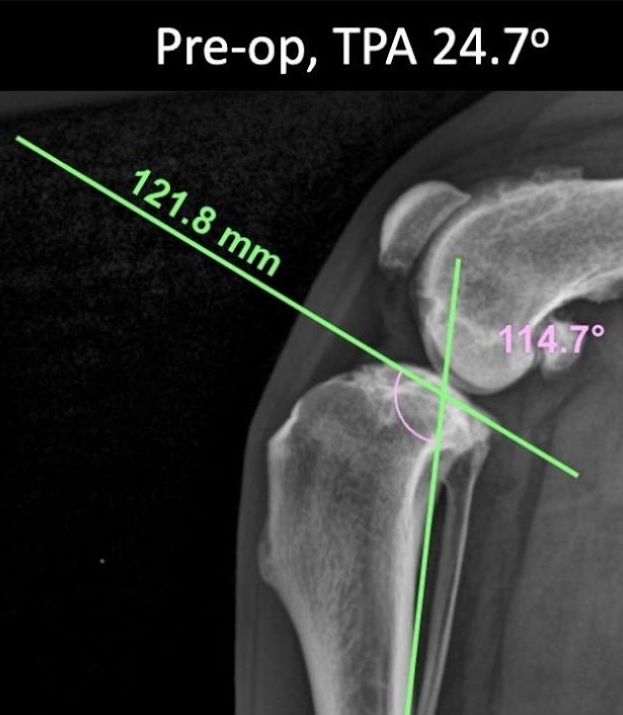
Digital Radiographs/X-rays
Radiographs are the next essential step in the process. We will evaluate the joint for swelling, assess the degree of arthritis (if any) present and use the measurements obtained by these x-rays to plan for surgery. Light sedation is all that’s needed for most dogs to obtain accurate x-rays and optimise the success of surgery. The hips and other knee will also be evaluated for evidence of arthritis or other conditions. A thorough re-examination of the movements of the knee will be repeated during this phase.
Advanced Imaging (Only in rare cases)
Ultrasound, MRi, CT are rarely indicated if the x-rays show evidence of other disease processes.
Treatment Options for Cruciate Rupture in Dogs
Surgery remains the gold-standard treatment to restore stability and slow arthritis progression in most dogs. Whilst non-surgical options are available and recommended by some sources, they are not as successful in obtaining persistent improvements of function as compared with surgery. We will discuss and give advice on all the options available to help your dog recover to the best ability possible.
Non-Surgical Management: Braces, Rest, Medication, Physiotherapy
Best for very small dogs with low-activity, suffering from certain conditions, or when surgery is not possible. Includes:
- Strict rest + weight management
- Anti-inflammatory medications & joint supplements
- Physical rehabilitation & custom bracing
- Platelet-rich plasma (PRP) injections (can be used with surgery as well)
Surgical Repair of Cruciate Ligament Rupture at Family Vet
MOST RECOMMENDED
Tibial Plateau Levelling Osteotomies (TPLO, CCWO, CBLO)
The tibial plateau is angled in most dogs. The cruciate ligaments prevent slipping of the femur relative to the tibia. After the ligament ruptures the femur slips down the slope of the tibia, causing it to displace forward. The gold-standard procedure is to level the tibial plateau. The physics is akin to preventing a ball rolling down a table or plank by keeping it level. If the tibial plateau is more level whilst walking the forces in the knee change and the knee joint does not luxate or move anymore while the dog walks.
Briefly this is achieved by making a cut in the top of the tibia and rotating or changing the top of the tibia. This will change the slope of the joint, eliminating the need for the cranial cruciate ligament. A titanium or stainless-steel plate holds everything stable while the bone heals. The exact procedure applied to achieve this (TPLO vs CCWO vs CBLO) will depends on the age, conformation and tibial plateau angle of the dog.
- Excellent procedure for dogs of almost all sizes
- It consistently achieves a faster return to normal activity
- 85–95% of dogs will achieve a return to full function
Extra-capsular (Lateral Suture) Technique
This was the traditional way to stabilise the knee joint for many years. A strong suture is placed outside the joint to mimic the cruciate ligament. Recent improvements lead to the development of procedures such utilising bone tunnels, more naturally positioned attachment points and different types of suture material, of which the “Tight Rope Procedure” is the probably the best known. It is better performed in smaller dogs or when budget is a concern. Some dog’s recovery may be slower, progression of arthritis can be increased or there may be implant related issues afterwards. It is sometimes combined with other procedures in those knee joints that need added stability.
Recovery & Aftercare
Most dogs walk within days after surgery
Important Disclaimer This is general educational information only. Every dog is different — factors like age, size, breed, concurrent issues (e.g., meniscus damage, bilateral disease), and individual healing vary. Always follow the exact protocol provided by your Family Vet surgeon. Do not start or change exercises without their approval. Over-activity can cause implant failure, delayed healing, or re-injury. Schedule rechecks (suture removal ~10–14 days; 8-week X-rays for bone healing).
General Principles of Cruciate Surgery Rehabilitation
Aim: Initially the goal is to control pain and swelling. This is followed by a phase where we want to maintain the range of motion of the joint to prevent freezing. This movement range exercises also help to clear the joint fluid, and ‘bathe’ all the structures in the joint with joint fluid in the recovery phase. The strength rebuilding phase and learning new proprioception/balance comes next. It takes around 3 to 6 months for most dogs to return to normal function and activity where both legs have equal muscle strength and balance. Whilst surgery is the first step, the rehabilitation is just as important to ensure successful use of the operated leg.
Keys to success: Strict activity restriction in the first two weeks. Most dogs are kept in a cage or very small room covered in a non-slippery surface such as a baby play mat. The dog will wear a protective cone to prevent it from licking and infecting the wound. This is a critical requirement. Pain medication and passive range of motion (flexing, extending or cycling motions) are essential to maintain range of motion and help reduce pain. Cold/hot therapy is helpful. After this initial phase progression to other exercise and short leash walks for toileting will be done. We strongly advise professional physiotherapy and hydrotherapy. The use of body harness, especially the ‘Lift Em Up’, is essential in dogs over 15kg.
Timeline Overview: We recommend a strict two week cage rest after surgery. Then two months with leash walks only for 10 minutes twice per day. Complete bone healing takes around 8 – 12 weeks during which the surgical site is protected by the special plate and screws. Full recovery to normal activities and muscle rebuilding is often achieved at 4-6 months for moth dogs.
Monitoring: Wounds for discharge, redness, swelling or heat. If the dog suddenly refuse to use the leg, or stops eating contact us immediately. STRICTLY prevent any licking/chewing in the first 2 weeks.
Note: Studies have shown that dogs with a rehabilitation schedule that includes physiotherapy (at home and professional sessions) improves the range of motion and use of their legs, have a better function and walk better than dogs without this rehabilitation.
What Exactly to Expect After Surgery: Typical Week-by-Week Rehabilitation Protocol
Weeks 0–2 (Acute Phase: The focus is on Pain Control & Early Range of Movement)
- Strict crate/small room confinement with non-slippery floor (baby mat or PAWZ type booties) — no stairs, jumping, running, rough play, or free roaming.
- Short leash walks only for toileting (3–5×/day, 5 minutes max, slow pace, short leash — full body harness/sling essential)
- Pain management: As prescribed (We use multimodal pain control)
- Cold therapy: Ice packs (wrapped) on surgical site 10–15 min, 3–4×/day (first 3–5 days) to reduce swelling and increase comfort.
- Passive Range of Motion Excercises: 2–3×/day. Gently flex/extend stifle (knee) 10–20 reps while dog lies on side (surgical leg up). Stop if painful.
- Massage: Gentle around thigh/stifle to reduce swelling (after cold pack).
- No active exercises yet.
- Wearing a cone for 24 hours is essential to prevent surgical infections
Weeks 2–4 (Transition: Start of Weight-Bearing & Basic Strength)
- Continue crate rest but increase controlled leash walks: 3×/day, build to 10 minutes.
- Stop cold packs; switch to warm packs before doing passive range of motion (PROM) exercises (10–15 min) to relax tissues and increase blood flow.
- PROM: Continue daily until the range of motion matches opposite leg.
- Introduce gentle home exercises (on non-slip surface, 5–10 reps, 1–2×/day):
- Sit-to-stand: Encourage slow sits/stands (use treats to lure).
- Cookie stretches: Lure head side-to-side/back for gentle stretches.
- Weight shifting: GENTLY rock dog side-to-side while standing.
- Three-legged standing: Lift non-surgical limbs briefly for balance.
- Ice after activity if sore.
- Recheck immediately if painful, stops eating, surgical site swelling or discharge
Weeks 5–8 (Strength-Building Phase: Muscle & Proprioception)
- Leash walks: 3×/day, build to 15–20 minutes. Add slow trots, gentle hills, or walking in long grass/water (low impact).
- Exercises (increase reps/time weekly):
- Sit-to-stand: Up to 15 reps, 2×/day.
- Cookie stretches & weight shifting: Add unstable surfaces (cushion, air mattress).
- Two-legged standing: Hold front or back legs up 20–30 seconds.
- Figure-8 weaves or ladder walks (if available) for coordination.
- Optional: Hydrotherapy (underwater treadmill) or professional sessions 1–2×/week — great for low-impact strength.
- 8-week recheck: X-rays to confirm bone healing → vet clears progression.
Weeks 8–12 (Advanced Strengthening & Return to Function)
- If X-rays good: Gradual off-leash in controlled areas (start 10–20 min/day).
- Add play: Tug (gentle, 5 min), swimming (build to 20 min).
- Continue strength exercises; focus on core/balance.
- Monitor for symmetry — opposite leg cruciate ligament tear risk is ~40–60% long-term.
Beyond 12 Weeks (Full Return to Activity)
- Gradual return to normal (runs, jumps, agility) over months.
- Maintain ideal weight, joint supplements, regular vet checks.
- Full muscle rebuild often takes 6 months.
Common Home Exercises to Help Regain Function and Strength
- Sit-to-Stand: Let your dog to sit then stand up slowly with treat — builds quads/hamstrings.
- Cookie Stretches: Hold treat at nose level, move side/side/up/back — improves neck/back flexibility.
- Weight Shifting: While standing, gently push hips side-to-side — improves proprioception. Do not push too hard to cause a stumble, gently swaying is the aim.
- Three/Two-Legged Standing: Lift opposite limbs briefly — challenges balance.
Additional Recommendations at Family Vet
- We can refer to certified canine rehab therapists for hydrotherapy, laser, TENS, or custom plans.
- Use non-slip mats at home to prevent slips.
- Track progress: Measure thigh circumference weekly; note lameness.
- Prevent opposite leg injury: Weight control + muscle building on both sides.
Why Choose Family Vet for CCL Surgery?
🔬
Orthopaedic Experience and Qualifications
Dr Wietz Botes – Registered specialist as a Diplomate of the American Board of Veterinary Practitioners in Canine/Feline with Master’s degree in surgery and extensive experience in surgery & minimally invasive techniques.
📍
State-of-the-Art Facilities
Digital X-ray, in-house laboratory, dedicated surgical suite in Fo Tan, Shatin.
❤️
Full Support
Pre- and post-op rehabilitation guidance, and compassionate family-focused care.
book 預約